Experiment#1:
Air We Breathe.
A close-up inspection of particulates in the air
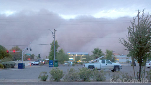
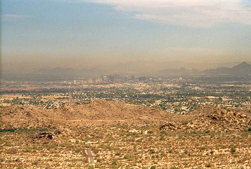
The image on the left is from one of the dust storms that hit Mesa and Phoenix. We all know that breathing that dust will be bad for us. Even on days without a dust storm, there are plenty of particulates in the air which aren't healthy.
Surprisingly, it's not just what the particulates are made of, but the size of the particulates that also affects its effect on the lungs.
Note: In this context, "particulates" mean particles that are suspended in the air.
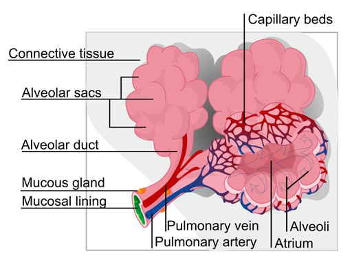
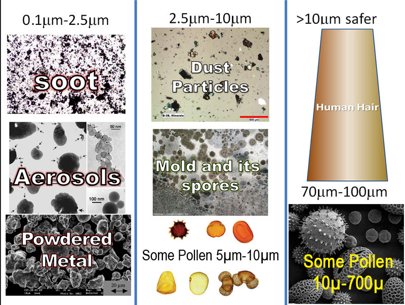

The air inside and outside our home contains numerous particulates (particles suspended in the air). Below are some, plus their size in microns. Anything under 10 microns is not visible to the eye and is more hazardous. By the way, dust mites live in your bedding and pillows. They eat the flakes of skin that you shed.
Human Hair .................(70 - 100 microns)
Pet Dander ..................(0.5 - 100 microns)
Pollen ...........................(5 - 700 microns)
Spores from Plants ....... (6 - 100 microns)
Mold........................... (2 - 20 microns)
Smoke ........................ (.01 - 1 micron)
Dust Mite Debris ........ (0.5 - 50 microns)
Household Dust .......... (.05 - 100 microns)
Skin Flakes ...................(0.4 - 10 microns)
Bacteria....................... (0.35 - 10 microns)
As you can see, all of these dip down into the unhealthy range of 0.1µm to 10µm.
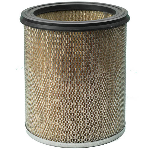
As a defense, filters are employed to trap these particles. You may have heard of HEPA filters for vacuum cleaners and room air filters. HEPA stands for "High Efficiency Particulate Arresting". Arresting meaning stopping. These filters were first used to capture radioactive particles in the air at the facility where the first atomic bomb was being built.
HEPA filters will only miss 3 out 10,000 particles 0.3 microns in size. For larger size particles, it misses even fewer of them.
It uses a mesh of fibers to accomplish this. Fibers can be made from plastic, glass, or cellulose (like cotton fibers).
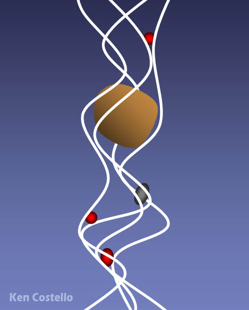
The fibers actually have openings much larger than the particles that it can capture. So it doesn't act like a seive that can only capture particles larger than the seive openings. As particles move through the fiber mesh, they get wedged in the narrow spaces where the fibers cross each other. The faster moving air actually helps particles get wedged in between fibers. That's probably why it is called High Efficiency Particulate Arresting. Of course, particles larger than the openings will get caught between the fibers.
They call these mechanical filters because the particulates are mechanically held (blocked or wedged) in the filter.
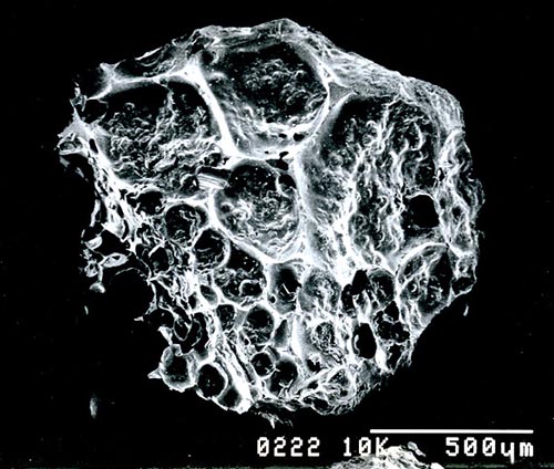
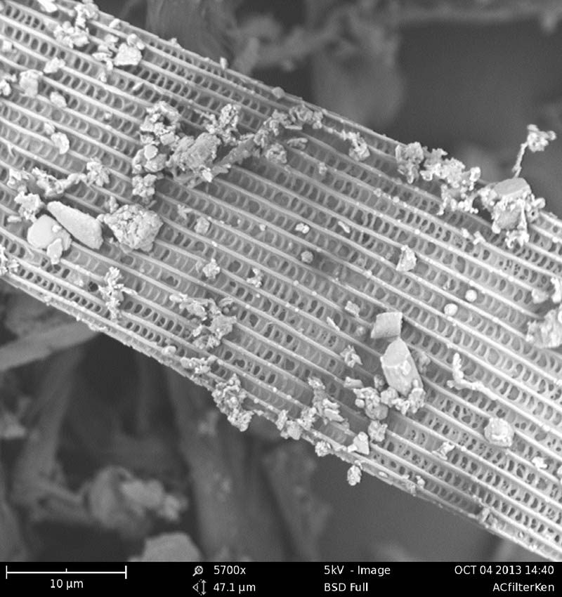
This is a granule of activated charcoal. The pits seen are about 100 microns in size; however, these are the large pits. There are others thousands of times smaller. So activated charcoal can act as a mechanical filter by blocking particles larger than its smallest pores. However, it can also trap certain substances that are so small that they can pass right through the pores. Activated charcoal does this because certain substance are attracted to the surface of the carbon. In other words, the carbon in activated charcoal acts like fly paper to certain substances. Vapors from solvents like gasoline or paint thinner stick to the surfaces and are trapped.
They call this adsorption (spelled with a "d"). Absorption (spelled with a "b") is when a thing soaks into something. Adsorption is when it sticks to its surface.
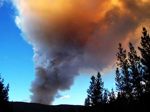
Smoke from wood fires:
The smoke from a campfire or from a forest fire is quite dangerous partially because of the abundance of very small particles that are in the smoke. Most particles in wood smoke are smaller than the 2.5 microns (2.5µm). These particles don't normally get coughed up. They are so small that they get stuck into the lining of the alveolus sacs.
erhaps the most dangerous component of wood smoke is the countless airborne particles (particulates). Airborne particles smaller than 2.5 microns are harmful because they are tiny enough to lodge inside lung tissue, while particles larger than 2.5 microns are coughed out. Researchers have discovered that the particles found in wood smoke are almost all smaller than 2.5 microns.
The Environmental Protection Agency lists the below health dangers for particulates:
- Irritation of the airways, coughing, and difficulty breathing
- Reduced lung function
- Aggravated asthma
- Chronic bronchitis
- Irregular heartbeat
- Nonfatal heart attacks
- Some cancers
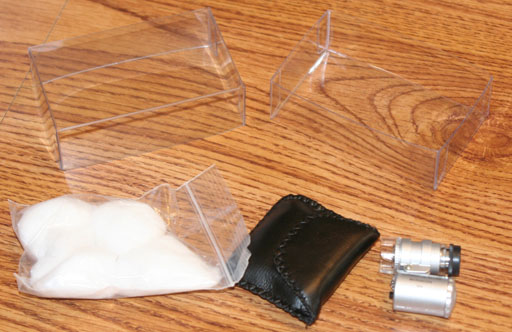
In your kit is a clear plastic case which contains a ziploc bag of cotton balls and a small black leather case, which contains the mini-microscope.
You also need some Scotch tape (invisible tape) for this lab. Recently, Scotch tape has been placed in the lab kit, but for most of the kits sold this semester, there is no Scotch tape included.
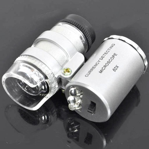
This lab uses the mini-microscope in your kit to see what particulates are in the air you breathe. The microscope is 60x, which is strong enough to see the particulates that have been discussed.
The left side of the microscope is the optics. The right side cylinder holds the batteries and the LED lights. It also has the small light switch to turn on the LEDs.
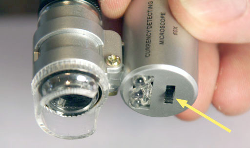
Sliding the light switch one way turns on a small ultraviolet LED light. Moving the switch the other way turns on two bright, bluish white LED lights. Use the bright LED lights to view particules you collect.
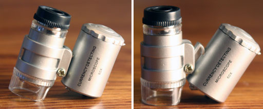
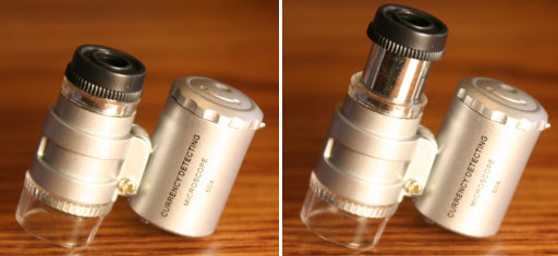
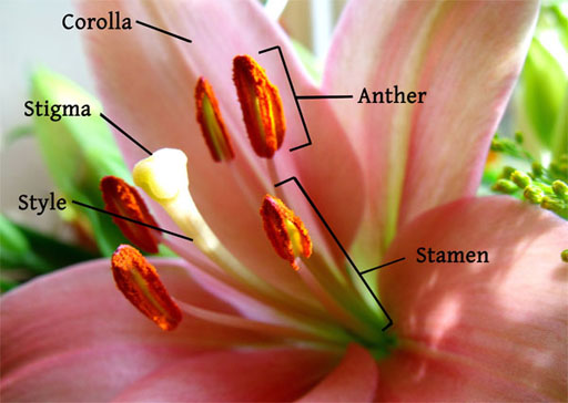
POLLEN: Before looking at a whole collection of particulates that settle out to the air, it's best to look at specific particulates, so that you can become acquainted with how they look. Pollen is one of the common particulates in the air. For people with allergies, pollen is one of the biggest triggers of allergic reactions.
Pollen is stored on the anthers, which sit on the ends of stalks called filaments.
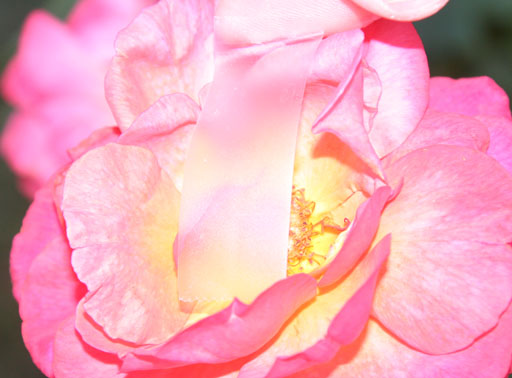
Your first task is to find something that is flowering. Using Scotch-tape, try to lift some pollen from the center of a flower.
If the Scotch-tape picks up some of the larger parts of the plant, just brush them off with your fingers. We are mostly interested in the grains of pollen.
 1. Take a photo of whatever flower you use to get a pollen sample. If you don't have a camera with you, you can take a photo of one of the other samples you will take later.
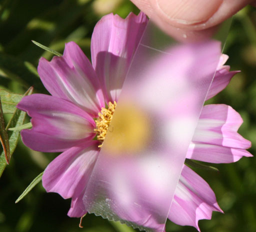
Here is another flower where we are using tape to lift up some of the pollen. You can use the same strip of tape if you want to get pollen from various flowers. Or you can just get pollen from one flower. Pollen grains are very small and often look like small yellowish particles.
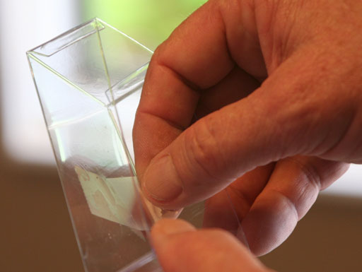
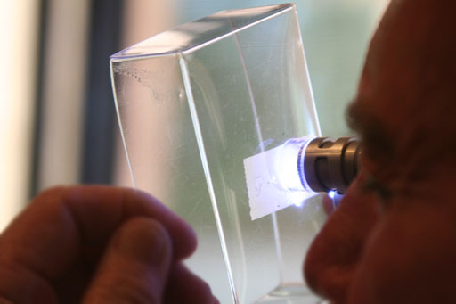
Turn the plastic case around so you are looking from the outside of the case. The tape is pressing the pollen up against the inside of the plastic case.
Hold your eye right up next to the eye piece. By lifting the microscope a little ways from the surface of the case, you can bring the pollen into focus.
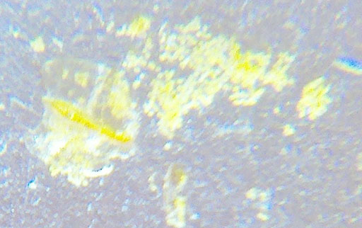
Depending on the particles, you sometimes see them better with a piece of white paper behind the Scotch tape. So have a piece of paper where you can place in the plastic box so that the paper sits behind the tape. The paper doesn't have to be right next to the tape but just behind it.
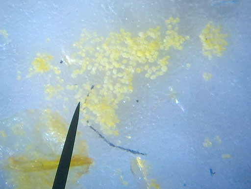
This is the same pollen taken with a more expensive microscope. We are still using a smartphone (Droid X). Like the photo above, this was taken with some white paper behind the pollen.
The pollen grains are the small round-like yellow granules. At least these are how rose pollen looks like.
The other pieces in the photo are plant parts from the flower.
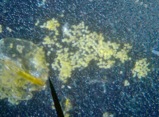
This is the same as the photo above but without the white paper behind the tape. The darker background helps the light colored pollen stand out. The small white specks that you see in the photo are just adhesive from the tape. So you have to learn to ignore the part of the image that is coming just from the adhesive.
2. Describe what your pollen looks like through your mini-scope.
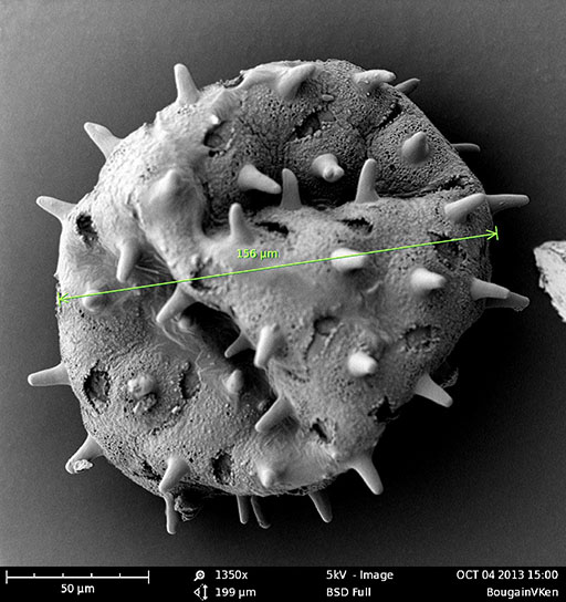
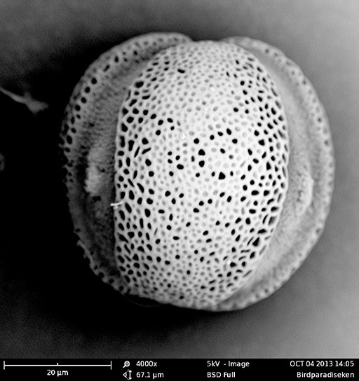
Here's another image I took. This pollen is from the Mexican Bird of Paradise plant. This one is much smaller than the one above. Notice the magnification is 4000x for this image. Using the 20µm scale at the bottom, we can estimate the size of this pollen to be about 60µm. That means this can get airborne and get into people's sinuses and lungs to cause an allergic reaction.
I like the pores on the surface of the pollen and the furrows. The number and orientation of the furrows is used to identify pollen.
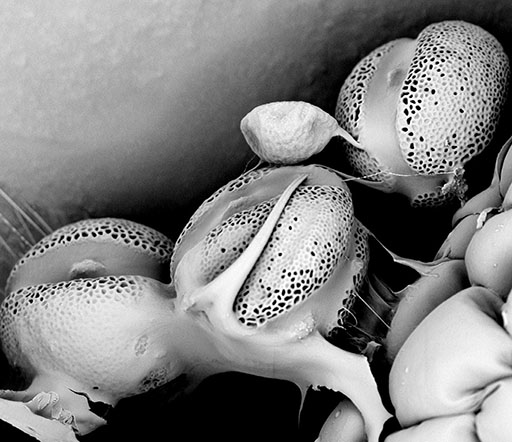
Here is the same pollen before it breaks free from the anthers. Note: Electron microscope images are only in grayscale. That doesn't mean what you see is gray. The pollen is probably yellow and somewhat transparent. Electron microscope imaging doesn't show transparent items as being clear because of the nature of the imaging and because the samples are coated first with gold.

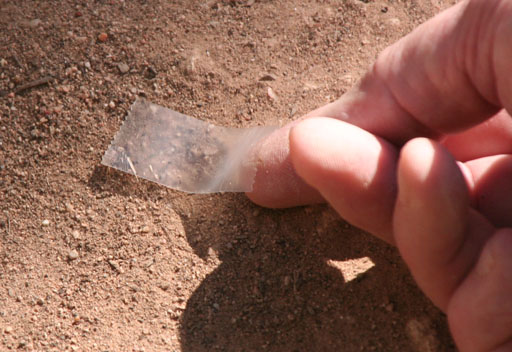
Find a patch of dirt and use Scotch tape to lift up some of the soil. If the tape picks up some larger pieces, brush those off with your fingers. We are mostly interested in the small particles in soil that can be lifted by wind.
 3. Take a photo of the area where you took the soil sample.

Just like you did with pollen, take the tape that you used to collect some soil and place it down on the inside of the plastic case that stored the microscope.
All samples you collect are mounted the same way. The are placed on the inside of the plastic box and then viewed from the other side.

Also, just like before, depending on the color of the particles, you sometimes see them better with a piece of white paper behind the Scotch tape.
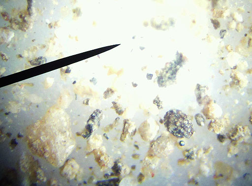
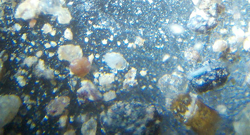
Here is the same sample without the white paper background. You can see that it changes the look of the particles. The lighter particles stand out more. The grains of minerals have a rough look. They also look clear, white, black, or may have some color in it. If it is a bright color, then it is probably not a mineral, perhaps a paint chip or piece of some insect.
4. Describe what you see with in your soil sample.
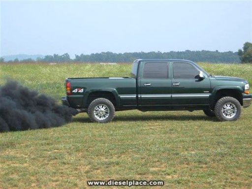
SOOT and SMOKE: The other particles that are often seen in air samples are carbon particles (soot). These come from unburnt gasoline and unburnt diesel. Fireplaces also put out a lot of soot, but fortunately in Phoenix fireplaces aren't used that much. Forest fires also put out a tremendous amount of ashes and soot. So it's just not the fire that is dangerous about a forest fire; it's the smoke.
When we think of diesel smoke, we usually think of the large semi trucks. However, there are thousands of pickups that have diesel engines just in any one city. The small trucks also put out diesel smoke. Not as much as the large trucks nor as much as the truck in this photo. Rather sadly, some diesel truck owners actually program their truck's microcontroller to produce these large amounts of smoke when they want to.

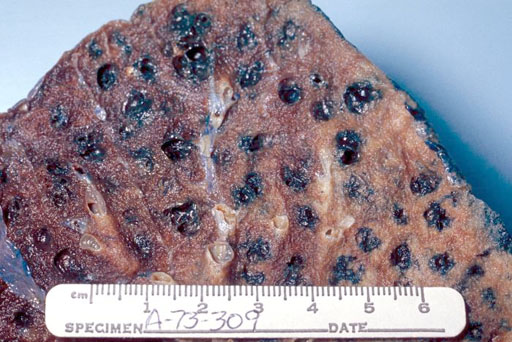
The ultimate air pollution happens when a person smokes. The smoke is deliberately inhaled deep into the lung. The very small particles of smoke penetrate into the lung tissue.
This is a section of lung tissue from a smoker. You can see how the carbon particles get lodged around the alveoli.
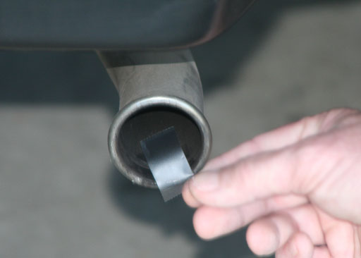
The inside of a tailpipe of a car is a good place to find some carbon particles. So use a strip of tape to take sample. Be sure the tail pipe is cool first.
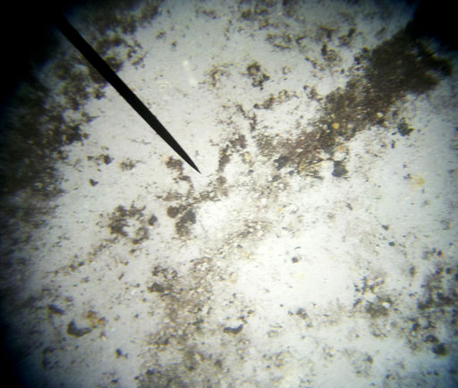
Here is the sample from the tail pipe.
Soot formed from unburnt gasoline or diesel are very small particles. They look like tiny black or gray specks under the microscope. In extreme magnification, they look like spheres. The reddish color in this image is probably from rust particles (iron oxides) coming from metal in the exhaust system.
4. Describe what you see in your sample from a car or trucks tail pipe. Also, say if it has a gas engine or diesel engine.
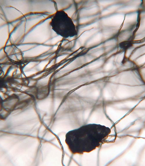
Some ash particles from cigarettes or burning wood can be larger than the typical soot particles. They are also black or dark gray. Some have a bluish tint. These ashes in the photo were caught in a wad of cotton using an air pump. The strands are the cotton fibers.
Again, the larger particles are normally caught by the nose or sinus. Unfortunately, smoking means the smoke goes directly into the mouth where this is little filtering. So it gets into the lungs without much stopping it.
FIBERS:
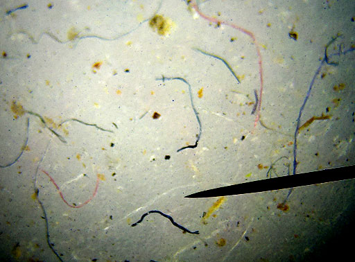
The air we breathe also has a lot of fibers and hairs in it. These short strands of fiber are from such things as clothes, rags, carpet, and paper towels. The hairs may be from pets or people, and even insects.

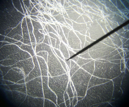
Cotton fibers look like flat ribbons that got twisted ribbons.
The light on your microscope is more bluish, so the fibers may appear a little blue than just white.
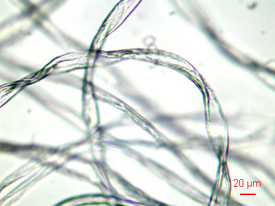
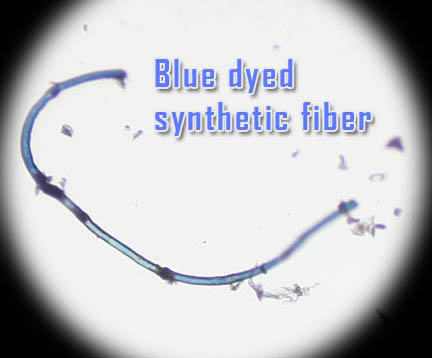
Synthetic fibers like nylon, polyester, and others are more round than cotton fibers. This magnification is about 100x so synthetic fibers in your scope will look thinner.
Long fibers don't get in the air easily. It's the short ones that we often breathe.

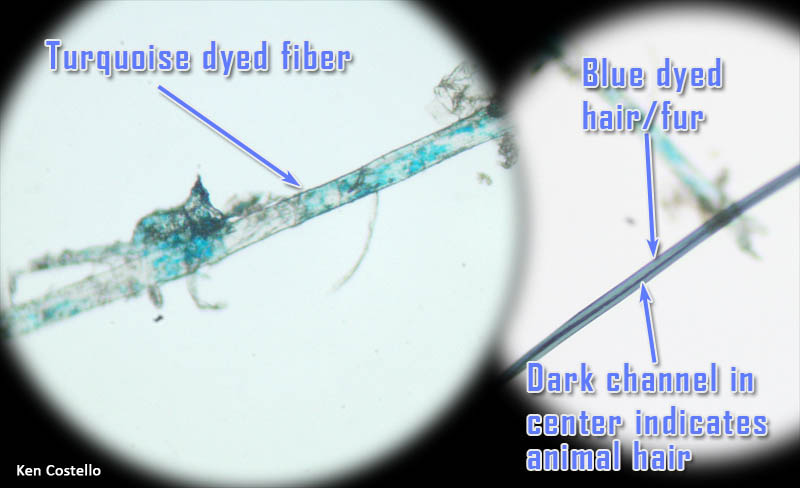
On the left is a synthetic fiber at about 250x magnification. Notice is it is not flat and twisted like cotton fiber. You can see some turquoise dye on (or in) the fiber. On the right is a strand of human or animal hair. Animal hair has a channel that runs through the middle of it. In high magnifications, scales on the outside of the hair can be seen..
DANDER AND DANDRUFF:
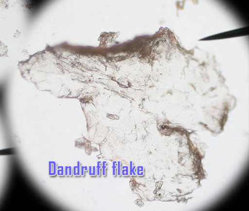
Both dander and dandruff are pieces of dead skin. Dander normally refers to the dead skin that comes off of animals like pet dogs and cats. Dandruff is dead skin from the scalp. Dandruff is usually larger and flatter than dander.
SAMPLE OF INDOOR PARTICLES:
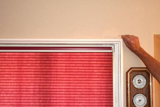
Now it is time to see the whole collection of what you breathe. Find a place that doesn't get dusted. That is usually the top of a door frame or window frame. Also, tops of refrigerators or other high places where it is hard to get to are good sources.
 Take a photo of yourself where you sampled the indoor dust. Make sure your face is visible. That proves you were there.
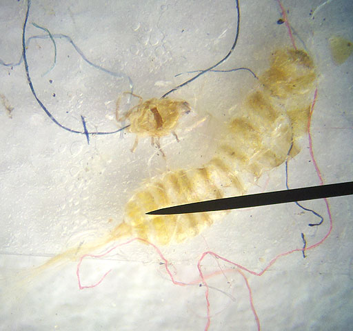
When I looked at the dust the tape picked up over a door frame, I was surprised to find two insects amid colored cotton fibers. The smaller insect looks like a dust mite, but its too big. I don't know what the big insect is. The black spots are likely soot.
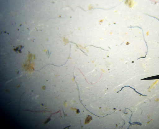
This is another portion of the tape from the top of a door frame. There are more fibers plus some black specks that could be soot or ashes. The brown particles are likely dander, plant pieices, and soil particles (dust).
5. Describe what you see in your sample that came from a place in the house that hadn't been dusted for a long time.
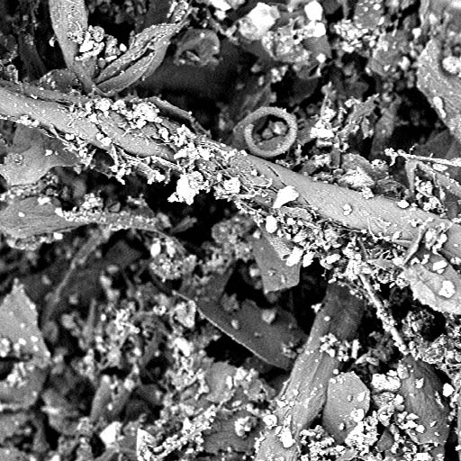
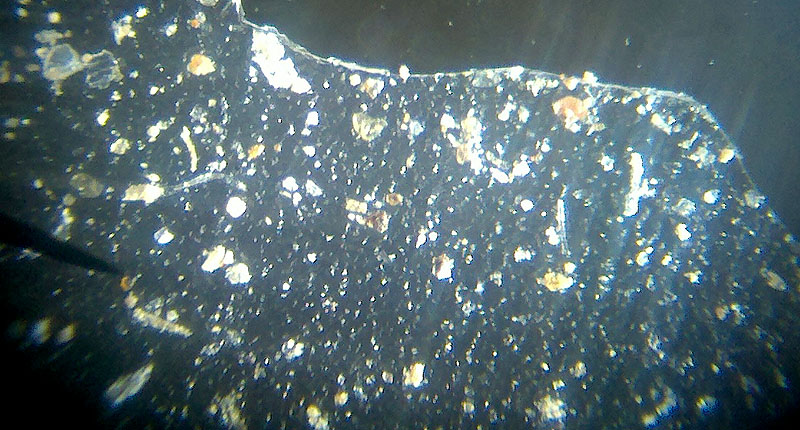
This is an image I took of some debris trapped in my A/C filter for my home. As you can see it is pile of different kinds of particles. The small white ones are likely grains of clay and quartz. The flat thin piece are likely flakes of dead skin. The tubular structures are fibers of some sort.
The magnification is 860x.
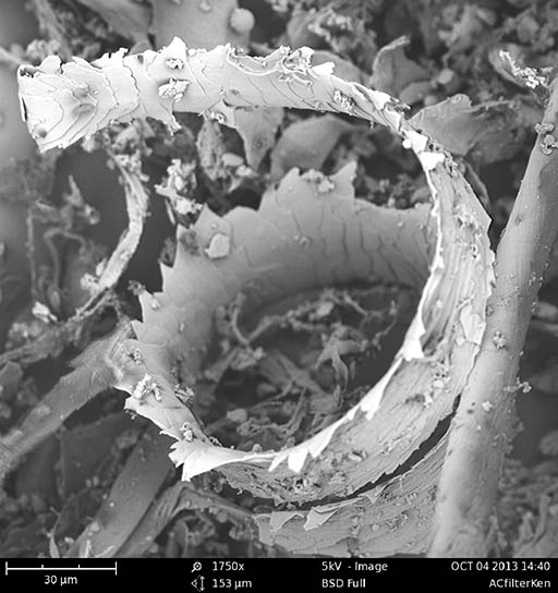
Take a sample from a dusty car or something else outside that is real dusty.
6. Describe what you see in your sample that came from outside that has accumulated a lot of dust.
Email your descriptions and photos to chm107@chemsitryland.com
